
QUINT / OBSERVATIONAL CASE STUDY
A ten-month Q.SCALP experience.
Explore one participant’s crown and frontal-hairline photographs, recorded routine and exploratory analysis over approximately ten months.
Explore the findings- Participant
- 30-year-old male
- Recorded course
- 7 applications
- Follow-up
- Approximately ten months
- Evidence
- One observational case
01 / WHAT WAS OBSERVED
A closer look at coverage.
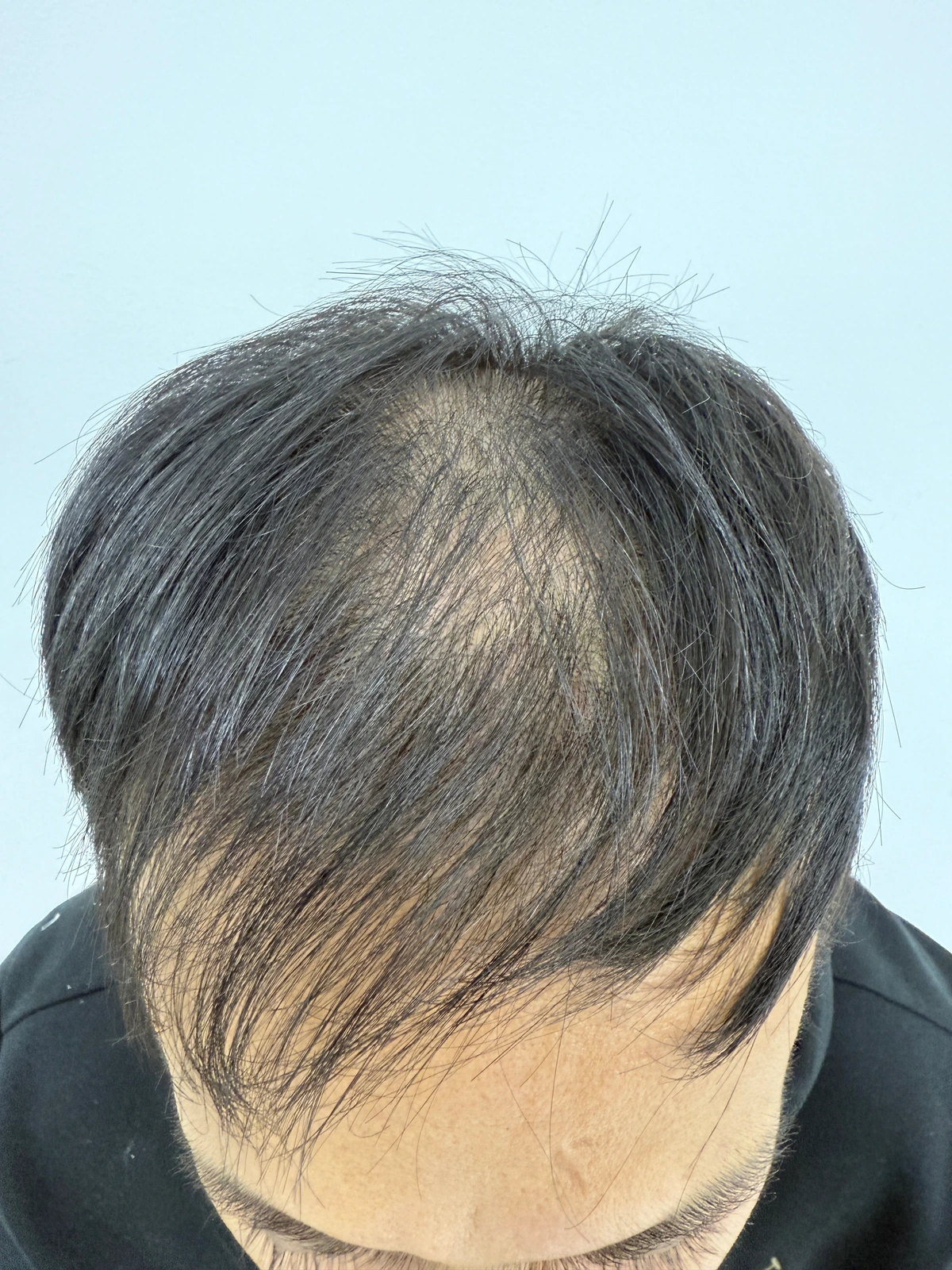
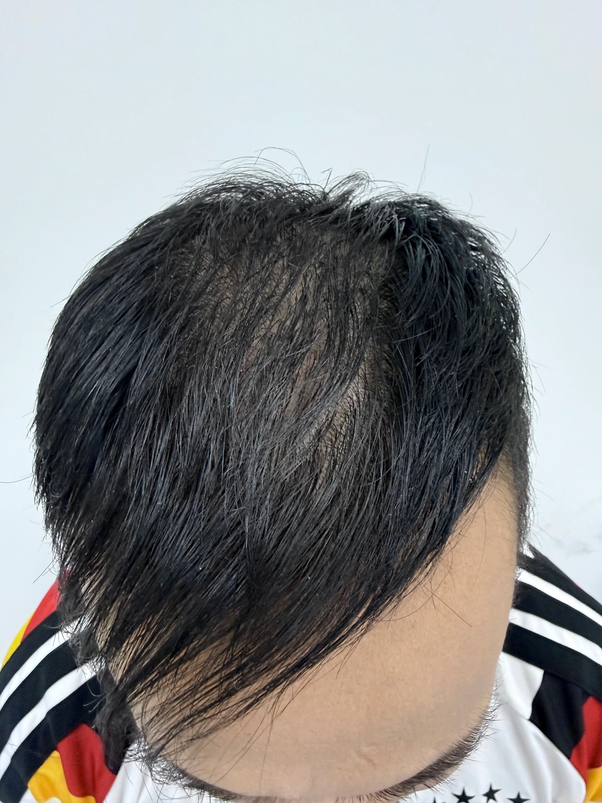
Photographs showed less scalp show-through in a selected central-crown area and a fuller-looking frontal fringe. Whole central-scalp findings were inconclusive.
The participant reported less shedding by month three, baby hairs by month six and a fuller look by month ten. These milestones were subjective, not independently measured outcomes.
02 / THE RECORDED ROUTINE
Seven applications. Ten months.
Four weekly Q.SCALP applications, followed by three approximately monthly applications. The final photographic record was taken about ten months after baseline.
- BASELINE
October 23, 2025
Overview photography before the recorded protocol.
- WEEKS 1–4
Four weekly applications
Q.SCALP applied to the crown and frontal hairline.
- NEXT 3 MONTHS
Three monthly applications
One application approximately every four weeks.
- MONTH 10
August 19, 2026
Overview, frontal fringe and baby-hair close-up photographs.
Seven applications exceed one three-session kit. This is the schedule documented for this participant. For current preparation, application and precautions, see the Q.SCALP product guide. View the Q.SCALP instructions.
03 / EXPLORATORY IMAGE ANALYSIS
Behind the numbers.
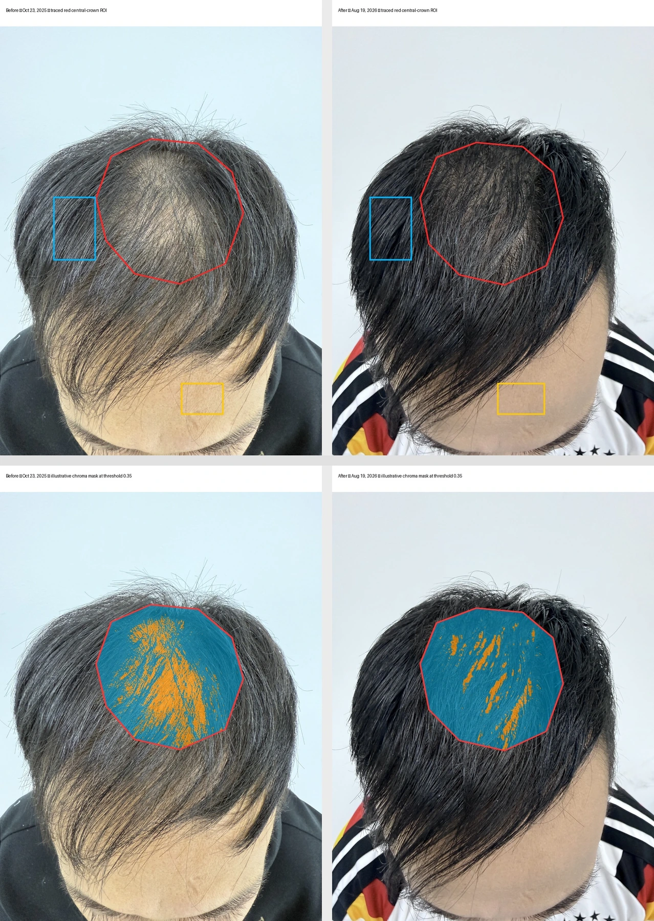
The strongest crown finding was localized to the selected red-outlined target. Broader regions and different thresholds give useful context.
Methods and analysis
SELECTED CENTRAL-CROWN TARGET
28.12% → 9.32%
Scalp-like color signal
At chroma threshold 0.35, the selected target’s scalp-like pixel fraction fell by 18.80 percentage points, a 66.86% relative reduction.
SELECTED FRONTAL FRINGE
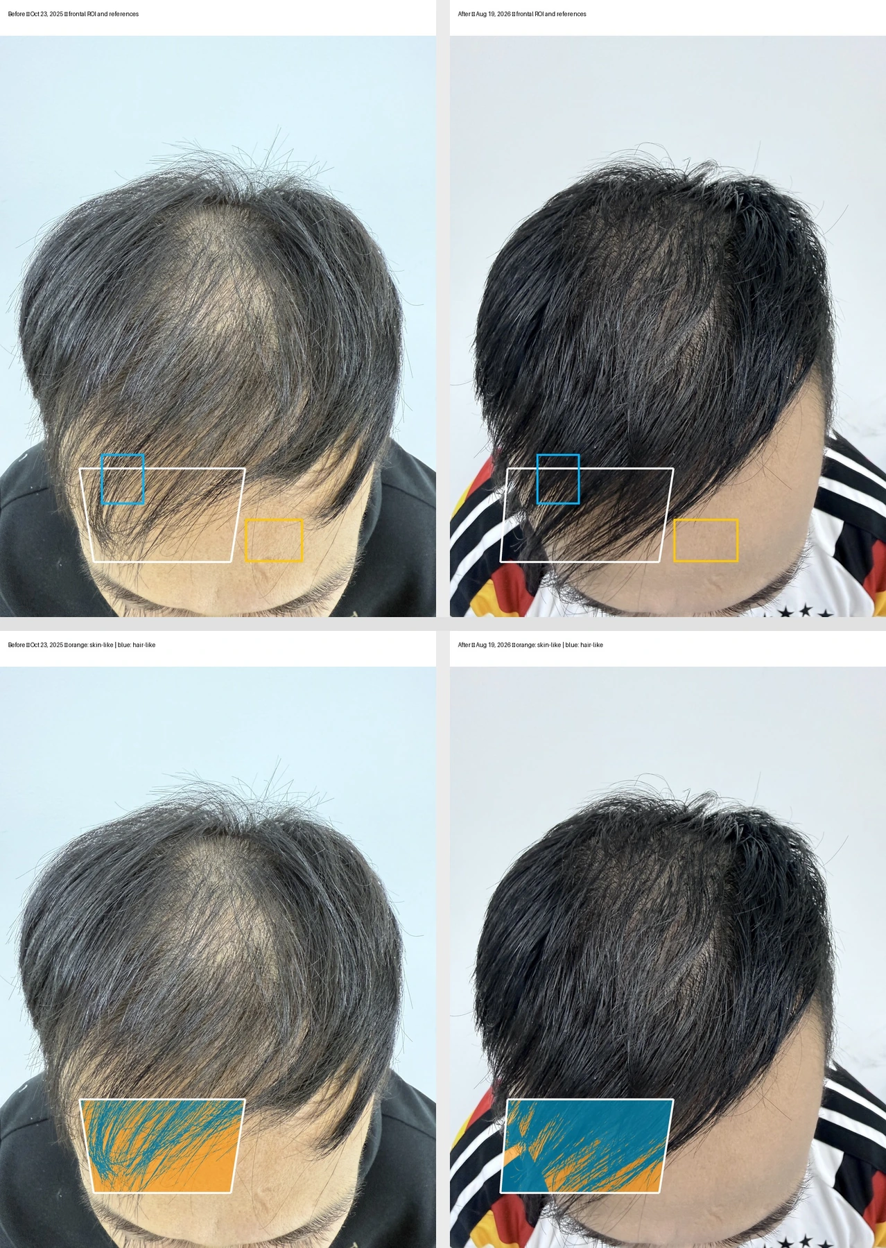
35.64% → 77.29%
Hair-like pixel coverage
The fraction of pixels classified as hair-like increased by 41.65 percentage points in the selected fringe region.
These values describe color and coverage within selected photographic regions. They do not measure biological hair growth, new follicles, hairs per square centimetre or individual hair thickness. The broader whole central-scalp measurement was inconclusive.
Crown findings: selected target and broader checks
The red target compares scalp-like and hair-like color while excluding brightness. Across thresholds 0.30–0.40, the relative reduction in scalp-like signal ranged from 57.70% to 77.55%. This is an analysis sensitivity range, not a hair-growth percentage.
| Region / measure | Baseline | Month 10 | Absolute change |
|---|---|---|---|
| Red target / threshold 0.30 | 39.34% | 16.64% | −22.70 percentage points |
| Red target / threshold 0.35 | 28.12% | 9.32% | −18.80 percentage points |
| Red target / threshold 0.40 | 16.39% | 3.68% | −12.71 percentage points |
| Broad upper target | 44.18% | 32.32% | −11.86 percentage points |
| Broad combined target | 39.64% | 40.76% | +1.12 percentage points |
The broader combined target did not show a reduction. Head angle and hair parting strongly affected the lower region. Red-target chroma and broad-target visible-scalp estimates are different measures.
Frontal fringe and the baby-hair close-up
Hair-like coverage increased from 35.64% to 77.29% in the selected frontal fringe and from 20.56% to 63.66% in the lower hanging-strand zone. Darker, broader overlapping bundles support a fuller-looking appearance; they do not establish thicker individual hair shafts.
The case team identified the short frontal hairs in the follow-up close-up as baby hairs. The selected field contained 17.60% hair-like pixels. No matched baseline close-up was available, so this is a follow-up observation rather than a measured increase in baby-hair count.
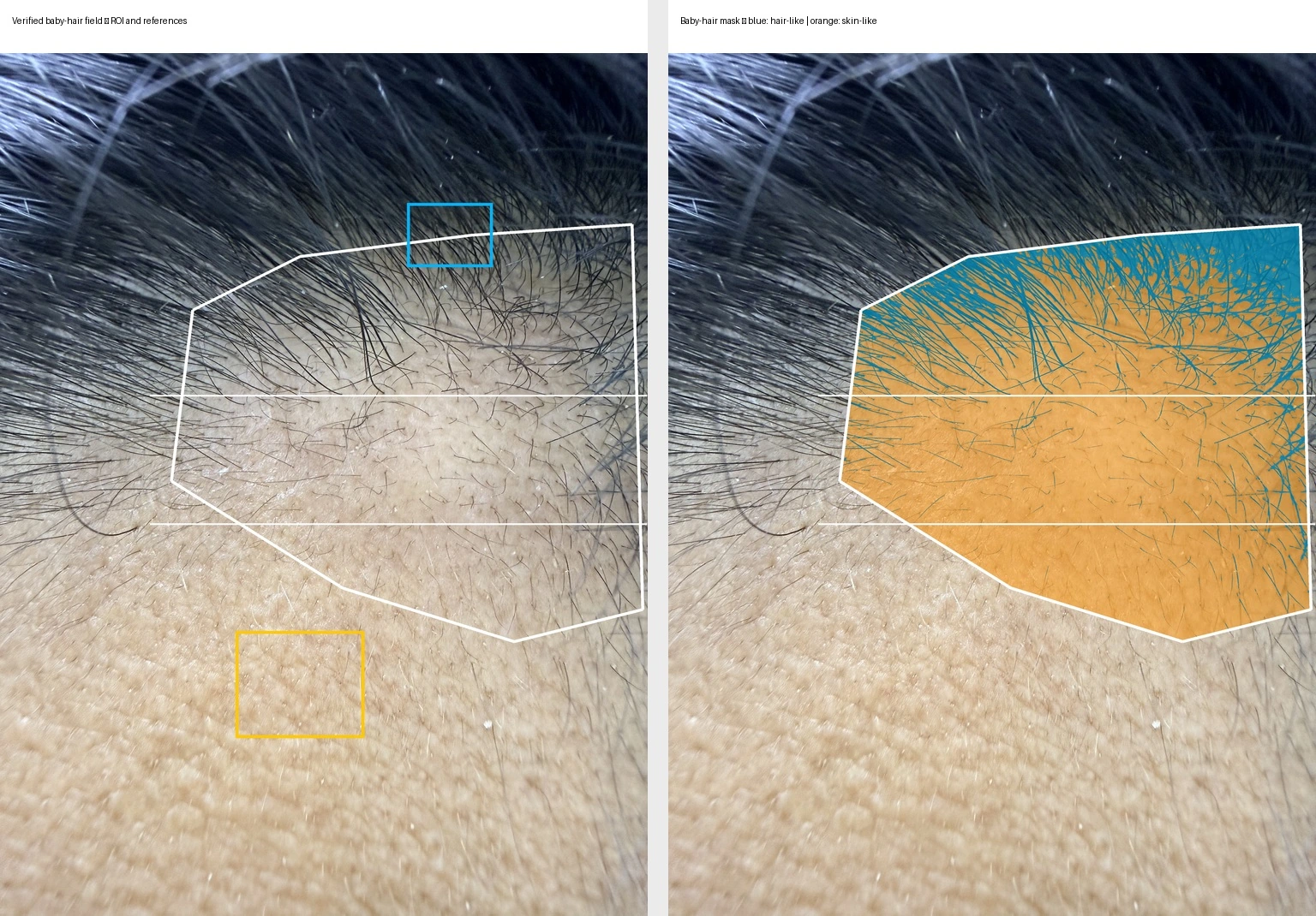
Image preparation and analysis method
The comparison at the top of this page uses the original photographs, without color-tone adjustment. The report’s analysis used high-resolution iPhone 16 Pro photographs, including a follow-up overview supplied with a global color-tone adjustment intended to better match baseline. Broad anatomical regions were manually matched because camera positions differed; the images were not registered to identical scalp tissue.
The central-crown method compared skin-versus-hair color without brightness to reduce darkness bias. Additional checks varied thresholds and region size. The supplied tone adjustment changed the broader RGB color-distance estimates by no more than 0.47 percentage points, but this does not remove other differences in the photographic conditions.
No inferential statistics were performed. Pixels and image regions are not independent participants. The full report includes the region definitions, masks and methods.
What this case can tell us.
The selected crown target and frontal fringe show localized changes in photographed appearance, alongside short frontal hairs documented at follow-up.
Single-participant observations. These photographs do not establish that QUINT caused the changes or predict typical results.
What remains uncertain.
- One participant, with no untreated control or blinded clinical assessment.
- Head angle, lighting, hair parting, clumping and apparent moisture or product state differed between photographs.
- Other treatments and lifestyle factors were not documented. There was no trichoscopy, physical hair-density measure or matched baseline baby-hair close-up.
THE COMPLETE RECORD
Read the full case study.
The supplied report, including methods, figures and limitations.
10-page report · PDF